Retinal Vein & Artery Occlusion
Retinal vein occlusion (RVO) is a blockage or occlusion of the retinal veins that carry blood away from the retina in the eye. This can lead to a buildup of pressure in the blood vessels, causing damage to the retina and resulting in vision loss.
There are two main types of RVO: central retinal vein occlusion (CRVO) and branch retinal vein occlusion (BRVO). In CRVO, the main vein that drains blood from the retina is blocked, while in BRVO, a smaller branch of the vein is blocked. CRVO is more severe and can cause more significant vision loss than BRVO.
The main risk factors for RVO include high blood pressure, diabetes, glaucoma, a history of blood clots or other vascular diseases, and age. Symptoms of RVO can include sudden vision loss, blurry or distorted vision, and the appearance of dark spots or floaters in the field of vision.
Retinal Vein & Artery Occlusion, BRVO, and CRVO Diagnosis
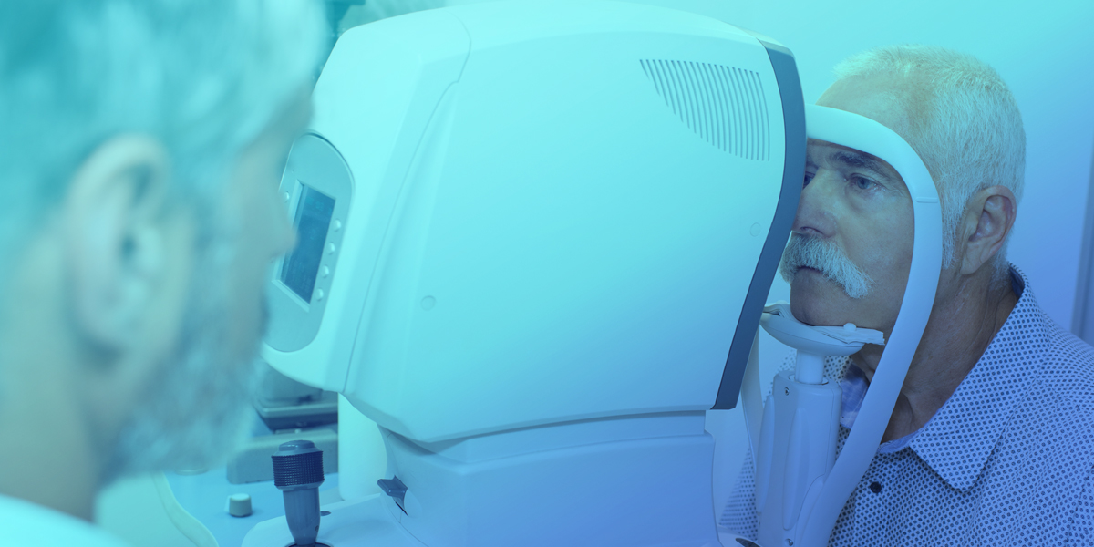
Regular eye exams, including a dilated eye exam and imaging tests such as fluorescein angiography or optical coherence tomography (OCT), can help detect RVO in its early stages, allowing for early intervention and treatment to prevent vision loss.
Treatment for Retinal Vein & Artery Occlusion
To protect your sight, it is important to recognize and treat a retinal vein occlusion as soon as possible.
Treatment for RVO depends on the severity and location of the occlusion and its complications. Macular edema, abnormal blood vessel growth, vitreous hemorrhage, or glaucoma may result. In most cases, intravitreal medications or laser therapy may be used to reduce swelling, promote blood flow, and restore vision.
The team at South Carolina Retina Institute works diligently to help patients preserve or improve their eyesight. Our aim is to provide the best care possible for your situation and we stay informed on the latest medical breakthroughs that show promising results.